


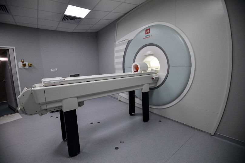
The French-built MRI - the most powerful scanner yet - is set to revolutionise the diagnosis and treatment of brain diseases, one pumpkin at a time.
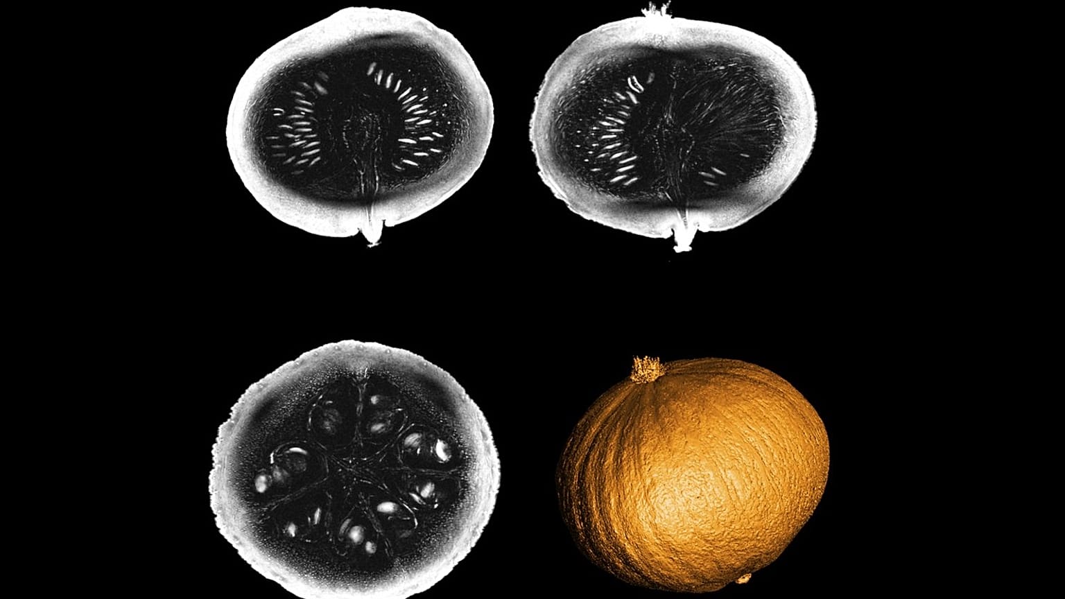
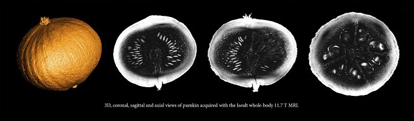
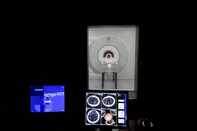
The world’s most powerful MRI scanner, which could have profound implications in diagnosing neurological diseases, has delivered its first images - of a pumpkin.
Called "Iseult," the machine is the fruit of more than 20 years of research and partnership between the French Atomic Energy Commission (CEA) and German manufacturer Siemens-Healthineers.
The machine will make it possible "to study the brain structure in a finer way," according to Cécile Lerman, engineer and project manager behind the new MRI scanner.
Why images of a pumpkin and not a human brain?
"It [the pumpkin] has the same diameter as the brain, an interesting, complex internal structure, with grains and fibrous parts - and then it’s the right season," Lerman said.
Before getting the green light from the health authorities for the examination of volunteer human subjects, the machine will first require months of careful adjustment and final tests of its instruments.
How does it work?
The machine doesn’t comply with ordinary standards. Its power surge peaks at 11.7 Tesla (T), compared with 3 T for usual examination machines. That’s the equivalent of 230,000 times that of the Earth's magnetic field.
Subjected to an intense magnetic field, the nuclei of molecules become polarised, much like a compass needle would be by the Earth's magnetic field.
The emission of weaker fields will upset this order and, depending on the frequency of the signal, make it possible to characterise and locate the type of tissues observed. Calculations then transform this data into images.
It took six years to manufacture the magnet and assemble the thousands of kilometres of a rare niobium-titanium alloy that make up the machine’s enormous 45-ton coil.
The magnet itself is maintained at a temperature close to absolute zero at -271 degrees Celsius, thanks to thousands of litres of liquid helium.
Such a process helps to remove any resistance to the flow of electric current in its coil.
The whole magnet, which weighs 132 tons, is housed in a cylinder 5 m long and 5 m in diameter.
"It’s a technological feat because we have pushed to their limits different techniques and materials, but also developed new calculation technologies" to produce images, Lionel Quettier, engineer and project manager for the magnetic part of the new MRI, explained to AFP.
The most efficient scanner for the best images
According to Quettier, Iseult is "the most efficient imager in the world".
It has become the new star of NeuroSpin, the brain imaging research centre of the CEA, located in Saclay near Paris, and directed by neuroscientist Stanislas Dehaene.
The centre already houses state-of-the-art machines, with MRIs at 7 T, and even 17 T that can accommodate small animals.
But Iseult will be the most powerful MRI capable of accommodating a human body thanks to its 90cm opening "tunnel".
The machine aims for an image resolution of under half a millimetre, a leap forward in the medical field that will allow "to go much more finely in the resolution of the spatial structures" of the brain.
The results could help to better understand the brain’s anatomy but also how it functions during cognitive tasks.
Neurodegenerative diseases such as Parkinson's and Alzheimer's, as well as psychiatric conditions like bipolar or vascular disorders could be prevented if spotted in time with this machine.