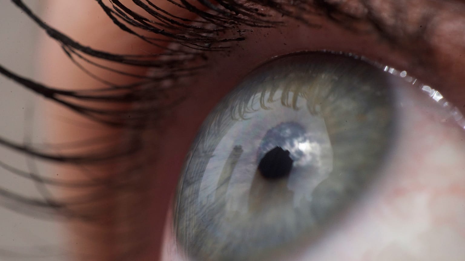
A 3D-printed, microscale implant in the eye that uses pancreatic cells could help to treat diabetes and other diseases.
Researchers in Sweden have developed a 3D-printed eye implant to offer cell-based therapy that could treat diabetes.
The device allows for pancreatic cells to be positioned in the eye and could make cell-based treatment for other diseases possible too, they say.
The researchers chose pancreatic islets, cell clusters in the pancreas that produce insulin, as donors’ cells have been used in experimental treatments for type 1 diabetes.
The immune systems of people with type 1 diabetes attack the cells that are used to make insulin – a hormone that plays a role in regulating blood sugar levels.
One of the problems with the experimental injections of donor pancreatic islets into people with type 1 diabetes is the need to prevent rejection of the cells.
The Swedish researchers say that implanting a device into the eye is thus ideal since it is "immune-privileged". This means that the eye can tolerate foreign matter or molecules without triggering an inflammatory immune response.
Donor transplants always have "a degree of reaction from the immune system," study author Anna Herland, who is also a senior lecturer in the Division of Bionanotechnology at Sweden's KTH Royal Institute of Technology, told Euronews Next.
"As the eye does not have resident immune cells we avoid these first reactions from the immune system," she added.
The eye’s transparency also allows researchers to monitor the implant over time.
Their research was published in the journal Advanced Materials this month.
'Mini organs' that integrate with the body
The device, which is about 240 micrometres long, was fixed between the iris and the cornea in the anterior chamber of the eye (ACE). It maintained its position for several months in tests on mice.
Wouter van der Wijngaart, a professor in the Division of Micro- and Nanosystems at KTH, said that they designed the device "to hold living mini-organs in a micro-cage and introduced the use of a flap door technique to avoid the need for additional fixation".
These "mini-organs" quickly integrated with the host animal's blood vessels and functioned normally, the researchers said.
Herland said their technology does not need invasive methods to monitor its function and guide care.
"Ours is a first step towards advanced medical microdevices that can both localise and monitor the function of cell grafts," she said in a statement.
It was the first "mechanical fixation" of a device in the eye’s anterior chamber.
While this was an early proof of principle study to determine if the concept could work, the researchers hope to advance the technology and that it may be used to treat other diseases.
"It can be used to host other organoid [tissue culture] like transplants. Today diseases are not treated by organoid like transplants except in trials with pancreatic cells but it is possible that in the future tissues like thyroid glands could be evaluated," Herland told Euronews Next.
This story has been updated to include quotes from one of the study's authors.


